Nearly 2,700 years ago, in an area in the present-day village of Heslington, UK, a middle-aged man was executed. Immediately after his death, his body began to decompose. Internal organs and flesh began to turn to mud. His hair turned to dust.
In 2008, archaeologists found his remains, with fragments of bones and skull remaining. Fractured cervical vertebrae confirmed the cause of death, he appeared to have been hanged. But when they looked inside the muddy skull, they suddenly saw a piece of brain tissue still intact.
Normally, the brain is one of the fastest decomposing organs of the body. Therefore, the piece of brain of the man in Heslington has become a big puzzle for more than a decade in science.
Why is the brain fragment of a man who lived from the Iron Age still intact until now, even though it has not been kept in a preservation environment?
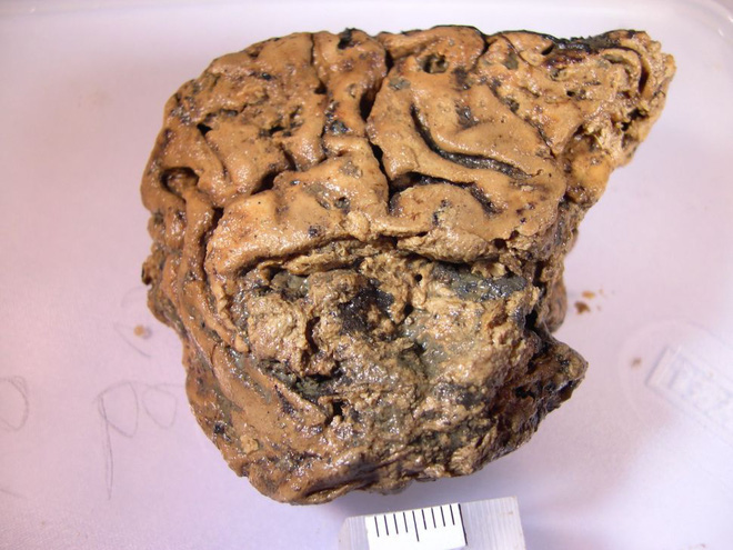
The mysterious piece of brain is nearly 2,700 years old, still intact even though it was not marinated
Exactly who this man was found in Heslington, and why he had to die, are perhaps questions that will never be answered. But analysis of the scene has helped archaeologists figure out some of the context of his death.
Carbon dating shows it was a middle-aged man. He breathed his last sometime between 673 and 482 BC. After being executed, probably hanged, his head was cut off and thrown into a hole. This hole contains a layer of fine sediment.
Normally, soft tissues in the body can only be preserved in three ways: dehumidification, freezing or keeping in an anaerobic, acidic environment. But this hole doesn’t have any of those conditions. As a result, other body parts, including the man’s hair, decomposed.
Only a piece of the man’s brain remained that amazed archaeologists, when they first saw it. The piece of brain was still intact and had the opaque yellow color of caramel. It looked like a small piece of tofu, only 80% the size of an adult human brain.
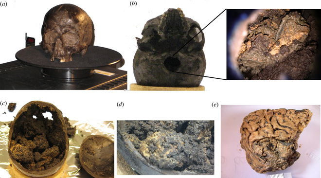
The skull that scientists found in the area of the village of Heslington, UK today
Taking over the sample and its puzzle that had existed for more than a decade, Associate Professor Axel Petzold, at the Institute of Neurology at University College London, spent months working, patiently analyzing the remaining protein samples. in the brain piece.
Finally, he has found the first clues to explain this amazing phenomenon of tissue preservation. ” The way this man was killed or buried may have led to the long-term preservation of his brain, ” Petzold said.
Before that, he had many years of experience researching two types of protein fibers in the brain: nerve fibers (neurofilaments) and glial acidic protein fibers (GFAP). Both types of fibers act like scaffolds, holding brain tissue structures together.
When Petzold and his team looked at the Heslington brain, they found that these filaments were still present in it, suggesting that it was the nerve fibers and GFAP fibers that contributed to the brain’s extraordinary preservation. Brain.
In most cases, a corpse’s brain will rot after enzymes from the environment and the deceased’s own microbiome eat and digest the brain tissue.
But tests that Petzold performed showed that these enzymes were deactivated within 3 months. That’s enough time for the proteins in the brain fragment in Heslington to automatically fold into a stable structure.
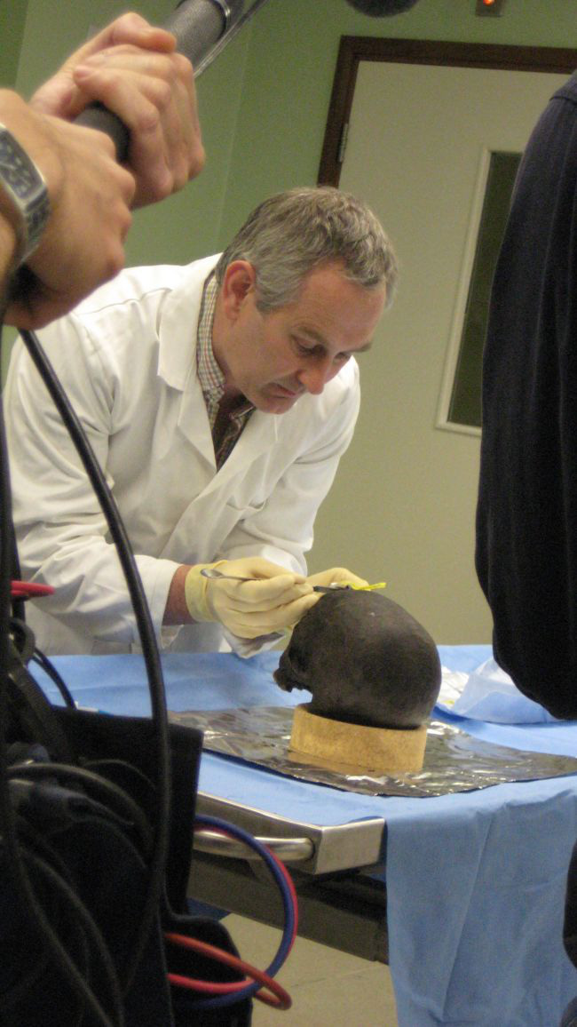
Something had seeped through the skull and preserved this brain.
Perhaps an acidic fluid invaded the brain and prevented these enzymes from chiseling just before or shortly after the person died, Petzold said. He gave more scenarios about this man’s death. Accordingly, in addition to the hypothesis of being hanged, he could have been beaten in the head or beheaded directly.
Normally, neurofilament protein is found in greater concentrations in white matter, which is located in the inner parts of the brain. But Heslington’s brain exhibits an abnormality, with more filaments in the outer areas, the gray matter.
Petzold suggests that a variety of factors may help prevent brain-degrading enzymes from starting in its outer regions, like an acidic solution seeping into the skull.
” The data suggest that the proteases [degrading enzymes] of the ancient brain may have been inhibited by an unidentified compound that diffused from outside the brain to deeper structures ,” he wrote in the paper research report.
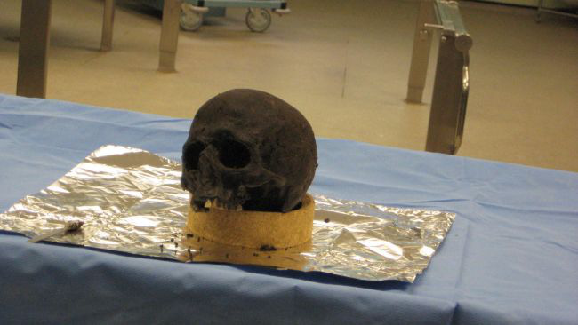
The mystery that the Heslington skull contains is not just a puzzle in itself.
In fact, the Heslington brain piece is not the oldest neural tissue sample ever found. Previously, archaeologists also unearthed smaller pieces of brain tissue, dating back up to 8,000 years inside a skull buried in water in Sweden.
But the mystery that the Heslington brain fragment hides in its excellent state of preservation is not just a puzzle in itself. Petzold’s findings about two types of protein fibers in the brain could also be the answer to a common neurological disease, Alzheimer’s.
The process of protein fiber clumping is also a neurological marker, a process that occurs in the brains of Alzheimer’s patients. To treat this disease, doctors will have to find a way to reverse that process. Petzold’s team looked at the protein clusters in the Heslington brain fragment and found that it took up to a year to unravel on its own.
This suggests that treatments for neurodegenerative diseases involving protein aggregation need to take a more long-term approach than previously thought.
The new study was published in the journal The Royal Society Interface
